
- Open architecture allowing the use of:
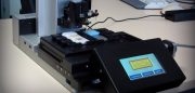
- 6 Standard Tissue Blocks mounted on “quick luck frame” on a MARZHAUSER (130x80mm) motorized stage.
- 3 Standard Tissue Blocks + 1 Macro Tissue Block mounted simultaneously
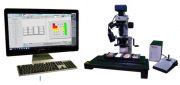
- 8 Mpixel Cmos Jenoptik camera
- Zoom 6000 Navitar Optics
- Wide Range of Standard Needles (from 0.6-1.0-1.5-2.0, 3.0 and 5.0 mm/Diam)
- All in one Computer (23.8 “ FHD: 1920 x 1080 Color Monitor)
- Windows 10 Pro Operating System
- Core selection (with manual or digital overlapping, with stretch function).
- Proprietary CK3600 SW (R) with “friendly user interface” allowing: TMA Geometry definition; TMA Design, TMA Construction and TMA reporting).
- Proprietary remote SW (allowing multiple users, to define the TMA geometry and Design at remote location.
- Generation of Excel files with the information related to each TMA core (e.g. Donor Block Identification Code, position taken from each Donor Block)
- Generation of XML files (with all core coordinates and related info) to feed to any commercial Digital Scanners (e.g. Aperio, Hamamatsu, TissueGnostics, Visiopharm, etc.) ensuring traceability, during the TMA Construction and Analysis of Tissue Core images.
- Possibility to construct Frozen Arrays by mounting the Galileo Frozen Module


